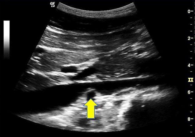
A 28-year-old male athlete underwent an abdominal scan. The emergency medicine ultrasound fellow obtained the following sagittal view of the mid-IVC.
What is the structure marked by the arrow?
A. Right renal vein
B. Right renal artery
C. Left renal artery
D. Left renal vein

The image shows the right renal artery
Explanation
The right renal artery is a branch of the abdominal aorta and it arises at level L1 or L2 below the origin of the superior mesenteric artery and the celiac axis. The right renal artery typically passes posterior to the IVC to reach the right kidney. There may be a situation where there may be an anomaly. The right renal artery may pass anterior to the IVC. Also, there could be multiple renal arteries. Surprisingly, multiple renal arteries are more common than expected. Multiple renal arteries are seen unilaterally in approximately 30% of patients and bilaterally in approximately 10% of patients. Presence of multiple renal arteries may be clinically significant in patients undergoing surgical or interventional radiological procedures involving the kidney.


