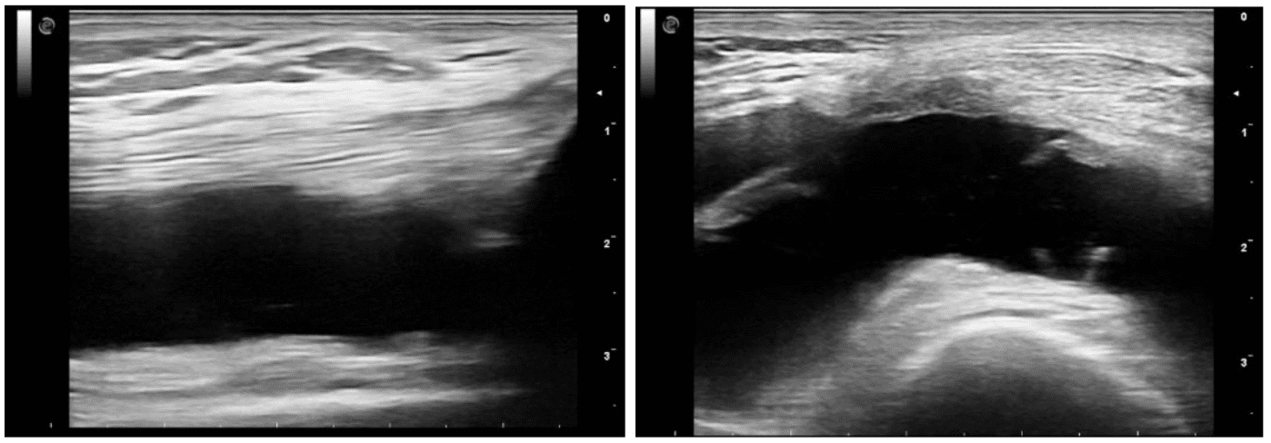
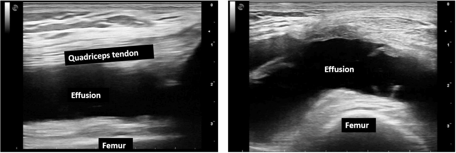
The following views were obtained in the suprapatellar region in the mid-longitudinal and transverse planes. There was no history of trauma or fever. The patient complained of swelling around the knee for the past 7 months.
What is the most likely diagnosis?
A. Hematoma
B. Abscess
C. Effusion

The most likely diagnosis is effusion.
Explanation
The mid longitudinal and transverse views shows a large anechoic collection in the suprapatellar recess region.

Figure – Mid longitudinal view (left) and suprapatellar transverse view (right) with a large anechoic (black) fluid collection.

Image courtesy of Wikipedia
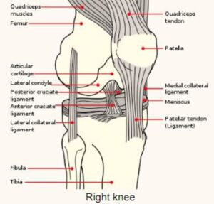
Figure – Right knee. Observe the location of the patella, quadriceps tendon and femur.
References
Test your knowledge of POCUS of Musculoskeletal Lower Extremity
with this knowledge check!


