By Victor Rao MBBS, DMRD, RDMS (APCA)
Introduction
Transabdominal ultrasound is a quick and easy modality to clinically assess a patient presenting with signs and symptoms that may suggest a pathology involving the kidney, ureter, or bladder. In this blog article we will review some common indications, basic scan techniques and tips as well as review some examples of commonly encountered urinary system pathology in the point of care setting. Do note that every possible renal pathology is not covered in this blog article.
Anatomy
The kidneys are retroperitoneal organs and normally lie on either side of the vertebral column at T12 to L3 level. Generally, the right kidney is slightly lower in position as compared to the left side due to the liver occupying the right upper quadrant.
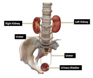
Figure 1. Normal anatomy of the urinary system. Observe the position of the kidneys, ureters, and bladder.
The adrenal glands are also located superior to the upper pole of the kidneys but are typically not well visualized on ultrasound in adults if they are not enlarged due to a pathology. The average length of an adult kidney is 10-13 cm. The left kidney is slightly longer in length as compared to the right kidney but not significantly different. Sometimes, one kidney may be shrunken or small due to some pathology. That will result in the normal kidney being significantly longer in length. Unilateral renal agenesis is present in 1:500 live births and the only kidney present is larger in size as compared to a normal adult kidney and those individuals should be advised to avoid contact sports as there is a higher risk of kidney injury. Bilateral renal agenesis also occurs rarely, and the incidence is approximately 1 in 4000 live births and is obviously incompatible with life and it can also be diagnosed during fetal ultrasound and should be suspected in cases where there is severe oligohydramnios or anhydramnios along with classic Potter syndrome. Renal agenesis may also be associated with chromosomal anomalies such as trisomy 21 and trisomy 22 etc. Do not confuse with ectopic kidney or crossed fused renal ectopia. If you are scanning an adult patient, it is impossible to be a case of bilateral renal agenesis. If kidney are not visualized bilaterally in their normal location, then scan entire abdomen and pelvic region to locate the ectopic kidneys.
Scan Tips
The kidneys should be scanned using a low frequency curvilinear/convex transducer with the patient in the supine position. If needed, the patient can be scanned in the left and right lateral decubitus position and even prone or sitting up. It is important to scan the entire kidney and bladder in both the longitudinal and transverse planes. Room lighting condition should be low illumination to avoid missing smaller renal masses or calculus. When in doubt about a small renal calculus use color Doppler to elicit the Twinkle artifact to enhance sensitivity of the ultrasound examination. Ideally the patient should be fasting overnight, and ultrasound examinations should be performed in the early morning hours to avoid excessive bowel gas. The patient can take sips of clear water if necessary and should be advised to hold urine to be able to assess the urinary bladder. The urinary bladder should be adequately distended but not overdistended. If urinary outflow obstruction is suspected, perform a bladder volume assessment pre and immediate post-void. Ideally, normal post void urine volume is zero. Since, urine is being produced constantly by the kidneys in a well hydrated patient with normal kidney function so we can expect 20-30 cc of urine in the bladder post-void.
Common Indications
Some common indications for renal ultrasound include flank pain, lower back pain, abdominal or pelvic pain, groin pain, hematuria, oliguria or anuria, urinary incontinence, nocturia, and dysuria. There could be some additional symptoms that may indicate the need to perform an urinary system ultrasound examination. This examination is relatively easy to perform once you master the technique.
Some Common Examples of Positive Findings
Renal Calculi/stones
Renal calculi or stones are commonly encountered during a KUB ultrasound examination. They appear hyperechoic on ultrasound. They could be an incidental finding in an asymptomatic patient. I remember a patient who had chronic lower back pain and was being treated with pain medication for several years.


