POCUS25: Top 25 Point-of-Care Ultrasound (POCUS) Community-Defined Practice Domains
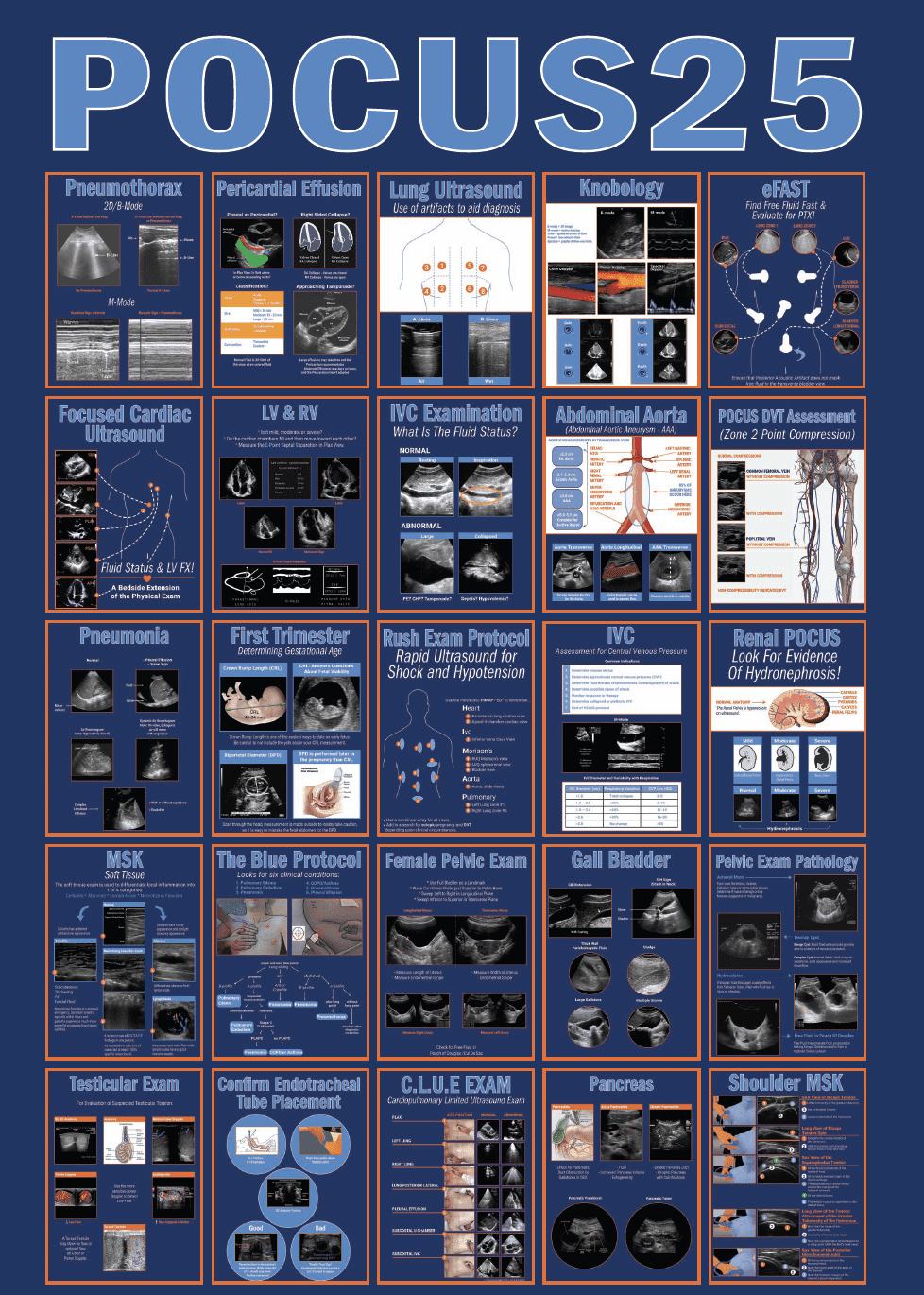
POCUS25 is a ranked list of the top 25 Point-of-Care Ultrasound (POCUS) practice domains, as determined by survey data obtained through the POCUS25 research study – a participatory longitudinal study launched in 2019 by the POCUS Certification Academy.
POCUS25 Practice Domains
1. Pneumothorax - B-mode and M-mode
Pneumothorax – B-mode and M-mode
Resources
- Infographic: Pneumothorax
- Webinars and Videos: Pulmonary POCUS (30 min)
- Case studies: Dyspnea – Lung ultrasound
- Blogs: Pneumothorax – can POCUS lung help? | Lung ultrasound vs chest x-rays: A brief overview
2. Pericardial effusion and cardiac tamponade detection …
Pericardial effusion and cardiac tamponade detection and differentiation from pleural effusion, assess IVC to support cardiac tamponade findings
Resources
- Infographic: Pericardial effusion
- Webinars and Videos: Cardiac tamponade (Podcast, 20 min) | Apical four chamber view of the heart (2 min)
- Case studies: Limited cardiac exam | MVA | POCUS apical 4 chamber view | Subcostal view of effusion
- Blogs: POCUS evaluation of pericardial effusion and tamponade
3. Normal lung ultrasound
Normal lung ultrasound – A-lines etc, lung sliding, lung edge, pleura and rib shadows, pneumonia/consolidation, air bronchogram, dynamic air bronchogram, spine sign
Resources
- Infographic: Lung ultrasound: Use of artifacts to aid diagnosis
- Webinars and Videos: Pulmonary POCUS (30 min) | Viewing lung A lines with POCUS (1 min) | Blue protocol: B-lines (1 min) | POCUS use in ICU (60 min) | Common pitfalls of lung POCUS in the ICU (1 min)
- Case studies: Lung ultrasound | Lung POCUS
- Blogs: POCUS lung – Introduction to A-lines and B-lines | Choosing POCUS can decrease radiation exposure in pneumonia patients | Lung ultrasound vs chest x-rays: A brief overview
4. Basic practical ultrasound physics and knobology
Basic practical ultrasound physics and knobology. Modes of ultrasound – B-mode/2D mode, M-mode, color Doppler, Power Doppler, spectral Doppler, depth, gain, TGC/DGC, common artifacts
Resources
- Infographic: Knobology | Mirror image artifact
- Webinars and Videos: 30-minute ultrasound physics university (35 min) | Doppler ultrasound demystified (70 min)
- Case studies: An ultrasound imaging artifact | Blood in urine | Gallbladder scan | Gallbladder sludge | Abdominal pain | Lung ultrasound
- Blogs: Renal Point of Care Ultrasound (POCUS) for nephrolithiasis
5. Protocol - EFAST
Protocol – EFAST
Resources
- Infographic: Abdominal blunt trauma
- Webinars and Videos: How using point-of-care ultrasound in emergency medical services (EMS) improves care (30 min)
- Case studies: Internal bleeding | A motor vehicle accident | MVA | eFast | Accident aftermath | POCUS eFast exam for chest and abdominal trauma | Abdominal trauma
- Blogs: Role of POCUS in abdominal trauma | POCUS: A first line of defense against time in the emergency department | Employing POCUS in CBRNE/WMD incidents | Honoring our veterans and the role of POCUS in the military | Role of POCUS in diagnosing ascites | Role of POCUS in assessment of the right upper quadrant to look for evidence of free fluid in the Morison’s pouch in a patient with abdominal trauma
6. Focused Cardiac ultrasound (FoCUS)*
Focused Cardiac ultrasound (FoCUS)* – PLAX, PSAX, A4C- ASE recommendation
Resources
- Infographic: Focused cardiac exam
- Webinars and Videos: Successful cardiac scanning (15 min) | Ultrasound use for vascular access and basic cardiac and pulmonary assessment for nurses (60 min) | Identifying false tendons in the left ventricle (1 min) | Basic point of care echocardiography views (60 min) | POCUS in cardiac arrest: The why, when, how and who (60 min)
- Case studies: Parasternal long axis (PLAX) view of the heart | Limited cardiac exam | Cardiac scanning
- Blogs: Left parasternal long axis/parasternal long axis (PLAX) view of the heart – brief review | POCUS for aortic stenosis
7. Gross estimation of LV and RV systolic function
Gross estimation of LV and RV systolic function – EPSS, contractility of LV and RV
Resources
- Infographic: Ejection fractions
- Webinars and Videos:
- Case studies: Left ventricular ejection fraction estimation | POCUS PLAX – Ejection fraction estimation
- Blogs: Right heart evaluation
8. Extremes of volume status
Extremes of volume status – hypovolemia and hypervolemia using IVC diameter and response to respiration and LV cavity diameter
Resources
- Infographic: Assessment for central venous pressure
- Webinars and Videos:
- Case studies: IVC: Upper abdomen view | Abdominal trauma
- Blogs: Inferior vena cava POCUS assessment | Inferior vena cava (IVC) assessment for volume status in POCUS setting | IVC ultrasound: Estimating fluid status & recognizing limitations | IVC Assessment Guide for POCUS Users
9. AAA screening, technique to measure ...
AAA screening, technique to measure diameter of abdominal aorta and aortic aneurysm, aortic aneurysm detection, thrombus in an aortic aneurysm and aortic dissection
Resources
- Infographic: Transverse aortic measurements
- Webinars and Videos: Screening for AAA with POCUS (30 min)
- Case studies: Fusiform aneurysm – case #1 | Fusiform aneurysm – case #2 | Fusiform abdominal aortic aneurysm | Abdominal aorta aneurysm (AAA) screening | Abdominal aortic scan | POCUS abdominal aortic aneurysm (AAA) screening | Abdominal aorta aneurysm screening
- Blogs: Patients who should be screened for abdominal aortic aneurysm | Abdominal aortic aneurysm | How to measure an abdominal aorta diameter/abdominal aortic aneurysm (AAA) diameter on ultrasound?
10. DVT
DVT – Lower extremity 2 zone compression test using B-mode only – saphenofemoral junction region and Popliteal
Resources
- Infographic: DVT assessment
- Webinars and Videos: Lower extremity venous assessment using ultrasound (2 point / 2 zone compression test) (30 min) | Anatomy and techniques for sonographers performing venous mapping (2 min) | Vascular applications of POCUS (60 min)
- Case studies: Pain IV site | Thrombus at the right saphenofemoral junction | Deep vein thrombosis (DVT)
- Blogs: Deep vein thrombosis (DVT): Is the blood clot always visible on B-mode ultrasound? | POCUS: Effective DVT diagnosis in immobile COVID patients | May-Thurner syndrome | Can we assess for a possible thrombus in the iliac veins with POCUS? | POCUS compression test for deep vein thrombosis assessment
11. Pneumonia/consolidation, air bronchogram ...
Pneumonia/consolidation, air bronchogram, dynamic air bronchogram, spine sign, pleural effusion, simple effusion, complex loculated effusion, anechoic and hyperechoic effusion
Resources
- Infographic: Pneumonia
- Webinars and Videos: Pulmonary POCUS (30 min)
- Case studies: Small pleural effusion | Lung ultrasound | POCUS eFAST exam | Subcostal view of effusion
- Blogs:
12. Basic first trimester ultrasound (TAS)
Basic first trimester ultrasound (TAS) – fetal number, fetal cardiac activity, amniotic fluid, intrauterine and extrauterine pregnancy, molar pregnancy, low lying placenta or placenta previa. TVS recommended for ectopic pregnancy if not detectable by TAS.
Resources
- Infographic: Determining intrauterine pregnancy in the first trimester
- Webinars and Videos: First trimester fetal biometry (60 min) | POCUS: A learnable skill (Podcast, 14 min) | Ectopic pregnancy (50 min) | Heterotopic pregnancy (1 min) | Introduction to transvaginal ultrasound (TVUS) (75 min)
- Case studies: Linear echogenic structure | POCUS in the late first trimester | Molar pregnancy | Lower abdominal pain
- Blogs: Molar pregnancy | POCUS: Managing the risks of ectopic pregnancies | Utilizing POCUS during early pregnancy to improve maternal health
13. Protocol - RUSH
Protocol – RUSH
Resources
- Infographic: RUSH exam protocol
- Webinars and Videos: How to differentiate various types of shock with POCUS (45 min) | Using POCUS to evaluate shock (2 min)
- Case studies: Abdominal trauma
- Blogs: RUSH protocol detects spontaneous retroperitoneal hemorrhage
14. IVC assessment for approximate CVP and volume status
IVC assessment for approximate CVP and volume status
Resources
- Infographic: Inferior vena cava exam
- Webinars and Videos:
- Case studies: IVC: Upper abdomen view | IVC anatomical challenge | Abdominal trauma
- Blogs: Inferior vena cava POCUS assessment | Inferior vena cava (IVC) assessment for volume status in POCUS setting | IVC ultrasound: Estimating fluid status & recognizing limitations | IVC Assessment Guide for POCUS Users
15. KUB ultrasound ...
KUB ultrasound, renal length measurement, renal calculi, hydronephrosis detection and grading, renal mass, ureteric obstruction, bladder volume, bladder mass
Resources
- Infographic: Renal POCUS
- Webinars and Videos: Introduction to renal POCUS (90 min) | The twinkle artifact (2 min) | Identifying horseshoe kidney (2 min)
- Case studies: Moderate hydronephrosis | Blood in urine | Colicky abdominal pain
- Blogs: POCUS in acute kidney injuries | Kidney, ureter, bladder (KUB) POCUS with some examples of commonly encountered pathology in the point of care setting | Renal point-of-care ultrasound (POCUS) for nephrolithiasis | Roles of POCUS in grading of hydronephrosis – A simple approach | The role of POCUS in the evaluation of suspected renal colic
16. Skin and soft tissue ultrasound ...
Skin and soft tissue ultrasound – normal appearance, edema, cellulitis, abscess
Resources
- Infographic: MSK soft tissue
- Webinars and Videos: MSK soft tissue ultrasound (65 min) | Baker’s cyst diagnosis (1 min)
- Case studies:
- Blogs: Can POCUS diagnose foreign body or air in soft tissue? | POCUS for skin and soft tissue infections (is it cellulitis or an abscess) – the basics | Dirofilariasis in humans: A rare POCUS spot diagnosis | Haglund’s deformity
17. Protocol - BLUE
Protocol – BLUE
Resources
- Infographic: BLUE protocol
- Webinars and Videos: The BLUE protocol: Bedside lung ultrasound in an emergency (35 min)
- Case studies:
- Blogs:
18. Basic female pelvic examination (TAS)
Basic female pelvic examination (TAS) – uterus, cervix, ovaries, adnexa, pouch of Douglas/cul-de-sac
Resources
- Infographic: Female pelvic exam
- Webinars and Videos:
- Case studies: Pelvic ultrasound | Transabdominal pelvic ultrasound
- Blogs: The midwife’s view with POCUS
19. Gallbladder ultrasound
Gallbladder ultrasound – gallstones, stone in the neck (SIN) sign, gallbladder mass, acute cholecystitis and chronic cholecystitis and gangrenous cholecystitis, ultrasound Murphy’s sign, CBD diameter and gross detection of CBD dilatation
Resources
- Infographic: Gall bladder assessment
- Webinars and Videos:
- Case studies: Gallbladder exam | Gallbladder scan | Gallbladder sludge
- Blogs: Role of POCUS in diagnosing acute cholecystitis and its life-threatening complications | Role of POCUS in diagnosing gallstones and some tips and important conditions to be aware of
20. Transabdominal female pelvic examination
Resources
- Infographic: Female pelvic exam
- Webinars and Videos: Role of ultrasound in diagnosis of ovarian cyst (60 min)
- Case studies: Pelvic ultrasound | Internal bleeding | An ultrasound imaging artifact | A transabdominal ultrasound | Lower abdominal pain
- Blogs:
21. Small parts
Small parts – testicular ultrasound, testicular mass, inguinal hernia, hydrocele, testicular torsion
Resources
- Infographic: Testicular exam
- Webinars and Videos: Spectrum of testicular pathologies of ultrasound (90 min) | Introduction to hydroceles (2 min)
- Case studies:
- Blogs:
22. To endotracheal tube position confirmation
To endotracheal tube position confirmation
Resources
- Infographic: Endotracheal intubation
- Webinars and Videos:
- Case studies:
- Blogs:
23. Protocol - CLUE
24. Pancreas ultrasound ...
Pancreas ultrasound, detection of pancreatitis, pancreatic mass, obstruction of CBD by a mass in the region of the head of pancreas or CBD calculus
Resources
- Infographic: Pancreas exam
- Webinars and Videos:
- Case studies: Pancreas assessment | Upper abdominal pain | Pain in the upper gastric region
- Blogs:
25. MSK
MSK – shoulder examination, basic view, ultrasound guided aspiration and injection, anisotropy
Resources
- Infographic: MSK shoulder assessment
- Webinars and Videos: Introduction to shoulder ultrasound (60 min) | An introduction to a knee ultrasound (60 min) | Percutaneous release of median nerve with PRP (30 min) | Dynamic evaluation of medial meniscus (1 min) | Posterior knee joint (1 min) | The 6-minute shoulder exam (60 min) | The 6-minute hand and wrist exam (60 min) | US-Guided PRP for Sports Medicine (60 min)
- Case studies: Elbow swelling
- Blogs: POCUS: the MVP of sports medicine | Ultrasound assessment of the long head of the biceps tendon (LHBT) – A brief overview
POCUS25 Research is Ongoing
POCUS25 is intended to be a living list that adapts to changing healthcare needs and practices. There’s still time to participate in future versions.
Contribute to POCUS25
We’re looking for your comments! Fill out the form to give us feedback on our POCUS25 research.


