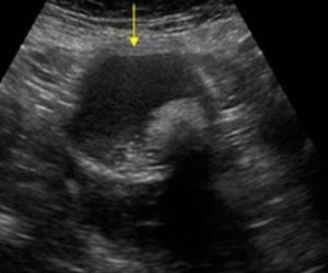
A 60-year-old female presented with vague lower abdominal pain present for the previous 6 months.
A transabdominal female pelvic ultrasound showed the image below.
What is the most likely diagnosis?
A. Distended urinary bladder with debris and large stone
B. Dermoid cyst
C. Endometrioma

Images courtesy of UltrasoundCases.info owned by SonoSkills
The most likely diagnosis is dermoid cyst.
Explanation
This is a classic diagnostic image of a Dermoid cyst. Never miss this diagnosis. The B-mode ultrasound image shows a complex cystic mass with hyperechoic components. The larger hyperechoic component also has a posterior acoustic shadow and is known as the iceberg sign. Carefully observe the echogenic dots and dashes next to the large echogenic structure. It is known as the dot-dash sign. This is due to hair inside the mass. Observe the yellow arrow pointing to the relatively hyperechoic fat floating on the top of the anechoic fluid inside the lesion. Note that the fat fluid interface is a straight line due to the fat floating on top of the fluid. These are all key features of a dermoid cyst, and this should be a spot diagnosis. You may sometimes see larger hyperechoic calcified lesions inside the cystic structure. Those would be suggestive of teeth inside the lesion (not seen in this lesion).
See the references below.
References
- https://doi.org/10.1177/8756479316664313
- https://radiopaedia.org/articles/mature-cystic-ovarian-teratoma-1?lang=us
Test your knowledge of Obstetrics First Trimester POCUS
with this knowledge check!


