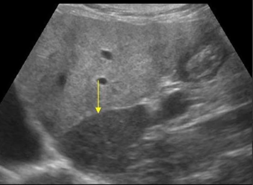
A 62-year-old obese male with elevated liver enzymes underwent a right upper quadrant POCUS exam. The liver showed fatty changes with a hypoechoic caudate lobe (yellow arrow).
What is the most likely diagnosis?
A. Liver metastases in caudate lobe
B. Liver abscess in caudate lobe
C. Fatty liver with focal sparing of the caudate lobe

Images courtesy of UltrasoundCases.info owned by SonoSkills
Explanation
Focal fat sparing areas may be seen sometimes in patients with fatty liver and appear normal in appearance on ultrasound as there is no fat deposition in those areas of the liver.
References
Test your knowledge of Hepatobiliary/Spleen POCUS
with this knowledge check!


