
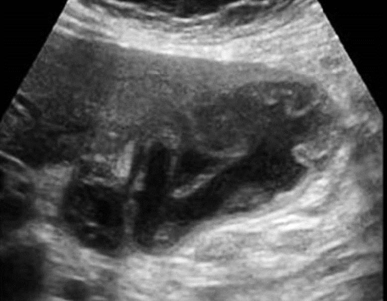
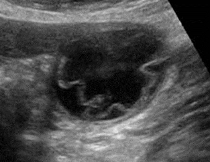
A 38-year-old female presented with pain in the right upper abdomen. The following two images were obtained while scanning her gallbladder.
What is the diagnosis?
A. Acute cholecystitis
B. Acute Gangrenous cholecystitis
C. Emphysematous cholecystitis
Images courtesy of UltrasoundCases.info owned by SonoSkills
This is a case of acute gangrenous cholecystitis.
Explanation
The images show a well distended gallbladder with folded membranous structure seen within the lumen of the gallbladder. This is the sloughed mucosal lining of the gallbladder. No air is seen within the gallbladder so it is not emphysematous cholecystitis. This is a surgical emergency and the patient must undergo urgent surgery to prevent a fatal outcome.
References
Test your knowledge of Hepatobiliary/Spleen POCUS
with this knowledge check!


