A 75-year-old female underwent abdominal ultrasound examination. The clinician observed that the IVC appeared suspicious. The following image and videos of the IVC in the upper abdomen were obtained.
What is the most likely diagnosis?
A. IVC tumor thrombus
B. No thrombus in the IVC – Rouleaux flow
C. Aortic dissection
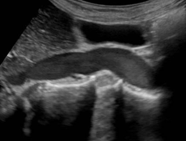
Image and videos courtesy of UltrasoundCases.info owned by SonoSkills.
No thrombus in the IVC – Rouleaux flow is the most likely diagnosis.
Explanation
The views obtained show the IVC lumen with a more echogenic appearance of the blood. On close examination of the video loops the blood in the IVC seems to be moving slowly towards the heart. The blood appears echogenic as the RBCs in the blood have clumped together and are now reflecting the ultrasound beam and thus making the blood appear more echogenic. This phenomenon is known as Rouleaux flow and is a reversible condition. The blood will become anechoic when normal flow velocity returns. The Rouleaux flow can be observed in the leg veins, IVC, heart chambers and IJV.
Do not confuse with a thrombus or DVT.
References


