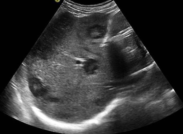
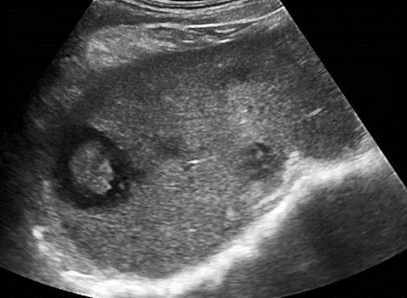
An ultrasound examination of the liver was performed on an 80-year-old male undergoing treatment for lung carcinoma. There was no history of fever or trauma. The following images were obtained.
What is the most probable diagnosis?
A. Liver abscess
B. Liver metastasis
C. Liver hemangioma

Image courtesy of UltrasoundCases.info owned by SonoSkills.

Image courtesy of UltrasoundCases.info owned by SonoSkills.
Explanation
The most probable diagnosis is liver metastases which may have variable appearance on ultrasound. In this case multiple hypoechoic lesions are seen within the liver parenchyma. The lesions have an echogenic center. Contrast enhanced ultrasound (CEUS) may also be helpful to visualize the lesion clearly. Biopsy is the gold standard.
References


