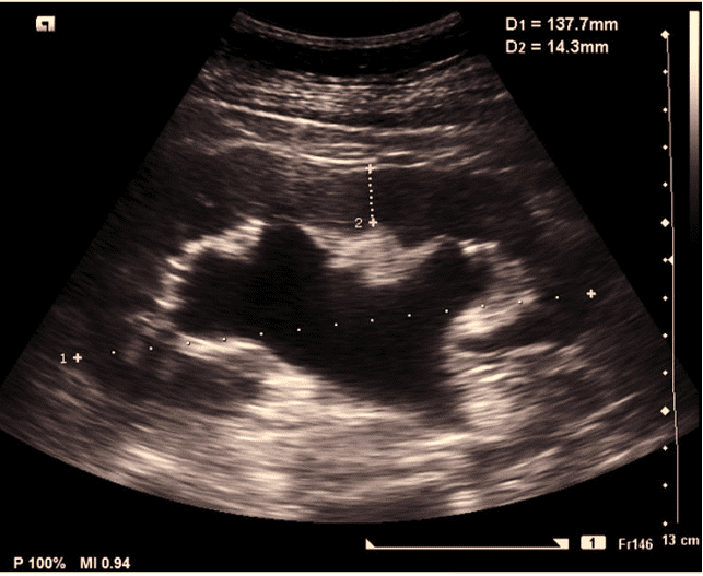
A 35-year-old female presented to the clinic with pain in the left flank.
A kidney, ureter bladder (KUB) was performed. The following image was obtained which shows moderate hydronephrosis. If this was a case of severe hydronephrosis, what would be the additional finding that would make a diagnosis of severe hydronephrosis?
The additional finding would be:
A. There would be evidence of a large staghorn calculus in the renal pelvicalyceal system.
B. The minor calyces would show enhanced concavity towards the periphery.
C. The renal parenchymal thickness would be significantly reduced.

Image courtesy of Radiopedia.org
Explanation
In a case of severe hydronephrosis we will see more obvious dilatation of the pelvicalyceal system.
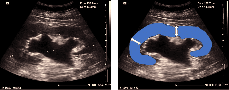
The important finding in severe hydronephrosis is renal parenchymal thickness reduction and in extreme cases could be even as thin as a membrane. Normal average renal parenchymal thickness is approximately 1.5 cm. If it is reduced to 1.0 cm or less that would be significant. You can also compare with the normal kidney on the opposite side when possible.

Figure. Renal parenchyma highlighted in the image on the right.
References


