A 52-year-old female presented to the emergency room with pain in the epigastric region for 3 days.
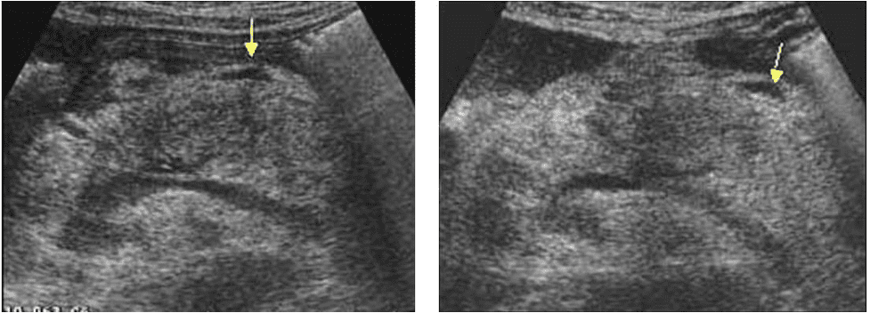
There was no history of trauma. Vitals were stable. A point-of-care ultrasound examination of the upper abdomen was performed. The following views of the pancreas were obtained.
What is the most likely diagnosis?
A. Pancreatic cancer
B. Paraaortic lymphadenopathy
C. Acute pancreatitis

Images courtesy of www.ultrasoundcases.info
The most likely diagnosis is acute pancreatitis.
Explanation
Ultrasound images show markedly enlarged pancreas with inhomogeneous echotexture and small amount of peripancreatic fluid (arrow). Ultrasound findings along with the history are consistent with acute pancreatitis. Serum amylase was 980 Units/Liter.

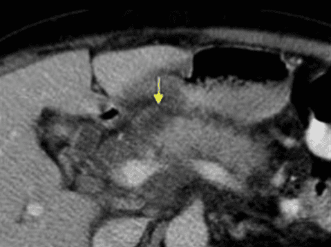
Fig 1. CT scan of the pancreas showing inhomogeneous edematous pancreas with small amount of peripancreatic fluid and some hypodense areas within the head, body and uncinate process of the pancreas.

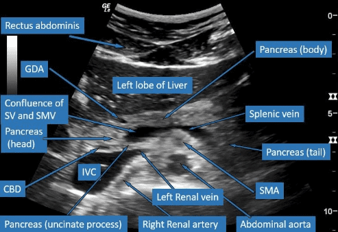
Fig 2. Labeled image of the pancreas showing the complex anatomy.
References


