A 35-year-old female underwent a transabdominal pelvic ultrasound examination.
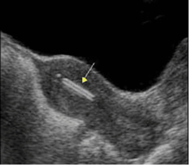
The image below shows a mid-longitudinal view of the uterus.
What structure is the arrow pointing to?
A. Retained products of conception
B. Blood clot
C. Intrauterine contraceptive device (IUCD)

Images courtesy of UltrasoundCases.info, owned by SonoSkills.
Explanation
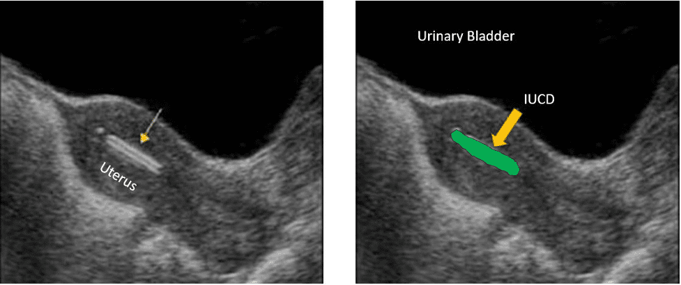
The image shows a linear hyperechoic structure in the uterine cavity. The finding is consistent with a properly placed intrauterine contraceptive device. Please see the references for a detailed explanation.

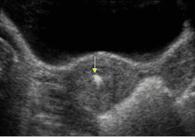
Figure 1. Transverse view through the uterus shows a bright/hyperechoic structure inside the uterine cavity. Ultrasound is commonly used to locate the position of IUCD.

Figure 2. IUCD inside uterine cavity
References


