By Victor V Rao MBBS, DMRD, RDMS
Hydronephrosis is defined as the dilatation of the major and or minor calyces and the renal pelvis due to an obstruction to the flow of urine from the pelvicalyceal system. It could be due to a calculus or an external compression on the ureter due to a mass or even due to urinary bladder outlet obstruction.
Point-of-Care Ultrasound (POCUS) is considered a sensitive modality to diagnose unilateral hydronephrosis. One study reported a sensitivity range of POCUS to be 72-83% to diagnose hydronephrosis as compared to CT scan. The study reported that 100% of patients with stones > 6 mm were diagnosed using POCUS. It is important to note that CT scan is the gold standard imaging modality to diagnose renal stones and hydronephrosis. However, that should be reserved for selective cases to avoid unnecessary exposure to ionizing radiation, higher cost and the increased time taken to perform the CT scan and obtain the results. POCUS has the advantage of low cost, availability at the point of care and no ionizing radiation. There are various methods of grading hydronephrosis. We will look at a simple approach to grade hydronephrosis in the point of care (POC) setting. Do note that there are various methods to grade hydronephrosis.
| Grade of Hydronephrosis | Ultrasound Findings |
| No hydronephrosis – Normal | A trace amount of urine may or may not be seen in the renal pelvis. Calyces – not significantly dilated or may not be dilated at all. |
| Mild hydronephrosis | Renal pelvis and calyces are dilated. Renal minor calyces are concave and do not balloon out towards the renal parenchyma or renal capsule. |
| Moderate hydronephrosis | The renal pelvis and calyces are dilated, and minor calyces are ballooned out towards the renal parenchyma or renal capsule and no longer concave in shape. Renal parenchymal thickness is preserved or slightly reduced. Normal renal parenchymal thickness is approximately 15 mm. |
| Severe hydronephrosis | The renal pelvis and calyces are more obviously dilated, and minor calyces are ballooned out towards the renal parenchyma and no longer concave in shape. Renal parenchymal thickness is reduced and could be very thin also suggesting damage to the renal parenchyma due to pressure within the renal pelvicalyceal system due to severe hydronephrosis. |
Table 1. One of the grading systems for grading hydronephrosis – may be helpful for POCUS users.
There is one drawback with this grading system. It does not differentiate between mild, moderate or severe reduction of renal parenchymal thickness. Do note that if the patient has moderate or severe hydronephrosis then the patient must be referred to a specialist and if presenting to the emergency department with moderate or severe hydronephrosis then the kidney specialist must be consulted, and a stent should be considered to bypass the obstruction and plan for corrective measures.
The patient may not fully understand the implications of the severity of hydronephrosis. Therefore, once we diagnose moderate or severe hydronephrosis it is the duty of the clinician to make sure that the concern is addressed appropriately. There have been instances when a patient presented with moderate hydronephrosis and then the patient did not follow up for few years. When the patient was seen after a few years, a diagnosis of severe hydronephrosis was made with complete destruction of the renal parenchyma and the patient had to undergo nephrectomy. This bad outcome was avoidable.
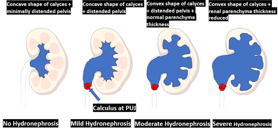
Figure 1. Diagrammatic representation showing the expected appearance of the pelvicalyceal system is different grades of hydronephrosis.
The image below shows a normal right kidney. Observe that the renal pelvicalyceal system is not dilated.
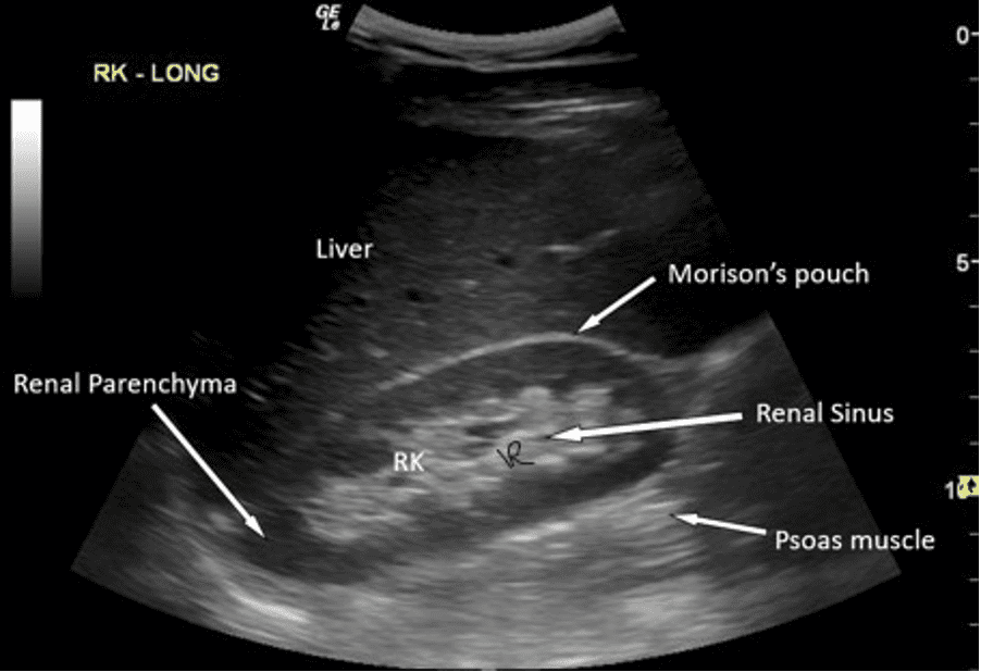
Figure 2. Normal right kidney with no evidence of hydronephrosis. Compare with mild hydronephrosis kidney below.
The image below shows an ultrasound image of the left kidney demonstrating mild hydronephrosis.
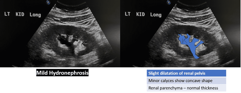
Figure 3. Mild hydronephrosis of left kidney.
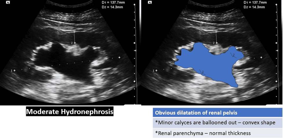
Figure 4. Moderate hydronephrosis. The minor calyces are blunted and more convex shaped. The renal parenchyma is almost normal.
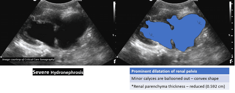
Figure 5. Severe hydronephrosis. The pelvicalyceal system is dilated with blunting of minor calyces. In addition, the renal parenchyma thickness is reduced. Renal parenchyma thickness is 0.592 cm in this case. Normal renal parenchyma thickness is approximately 1.5 cm.
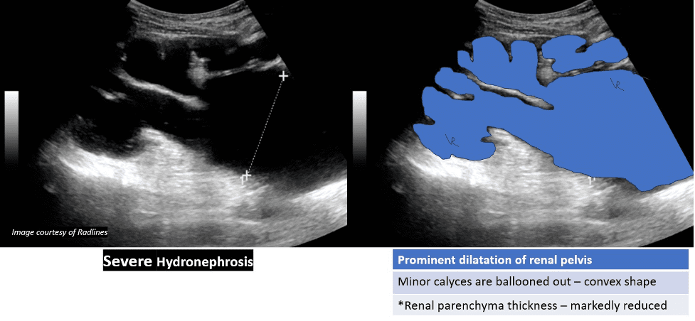
Figure 6. Another case of severe hydronephrosis. The pelvicalyceal system is more dilated than the above case. Note that renal parenchyma is markedly reduced. This would be consistent with more advanced stage and chronic severe hydronephrosis. In extreme cases the renal parenchyma is destroyed completely with no functional renal tissue.
References
https://www.ncbi.nlm.nih.gov/pmc/articles/PMC3935794/
https://www.frontiersin.org/articles/10.3389/fped.2020.00458/full
https://www.renalfellow.org/2019/05/10/the-ultrasound-mimics-of-hydronephrosis/
Access the full POCUS Learning Library for FREE!
Share a few details so we can tailor new content to your specialty and region.


